High-resolution Optical Micro-spectroscopy
Optical micro-spectroscopy deals with analyzing the properties of light scattered from a material to deduce information on its chemical and physical properties. The latter can be extracted by measuring the spectral or temporal changes in the light at very small and short intervals, respectively. These can provide information on collective molecular dynamics which in turn can be used to extract properties such as the microscopic viscoelastic properties - namely the viscosity and elastic moduli. When combined with a microscopy setup (such as a confocal microscope), this allows for non-invasive label-free 4D mapping of these parameters with near diffraction limited optical resolution (<500nm). While the microscope-design is often conceptually not too different from that of other scanning optical microscopes, the spectrometer design as well as analysis/interpretation are less trivial, and require the use of several “optical tricks” to achieve the needed sensitivity to deduce the parameters of interest.
Applications for anatomy: The morphology and function of our anatomy (and indeed that of any living system) across different scales is ultimately governed by the constituent physical (mechanical) properties. Understanding these are thus essential for understanding structure-function relations (e.g. why a certain anatomical feature looks the way it does, how and under what stresses or chemical conditions it changes, and how such changes will impact its functionality and affect the organism as a whole). Besides the fundamental anatomical insights that studying the microscopic mechanics can give us, numerous pathologies and the onset thereof are known to be associated with changes in the mechanical properties, both on the microscopic as well as on macroscopic scales. Studying these can be expected to give rise to conceptually novel none-invasive tools for prognostics, diagnostics, and medical quality-control (e.g. tissue engineering, transplants, etc.).
While the inventory is continuously evolving and changing, below is a list of setups that constitute our current infrastructure. In most cases the instruments have associated custom control, fitting and deconvolution software / scripts.
Instrument descriptions:
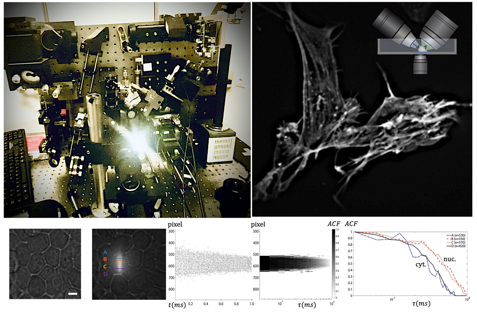
1. Confocal scanning Spontaneous Brillouin Light Scattering Microscopy.
Inverted microscope geometry (Olympus IX-73). Back-scattering geometry. Changeable objective lenses from 0.1 - 1.49 NA. Scan range: up to 300 microns (piezo stage) and 2.5cm (motor stage). Parallel correlative fluorescence and Raman scattering spectroscopy (with optional additional excitation at 405nm, 488nm and 642nm). Operating spectroscopy wavelength: 532nm. Elastic suppression via gas absorption cell, interference filters and spatial masks. Stable temperature control (room temperature to ~42C). Free Spectral Range: 30 GHz (60 GHz possible by replacing several optical components). Suitable for reasonably transparent solid and liquid samples. Also available is a hand-held fiber coupled probe that can be used in place of the microscope frame.
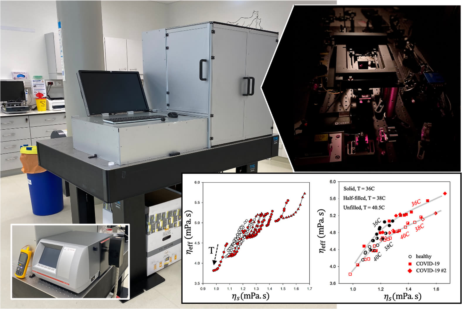
2. High-throughput Spontaneous Brillouin Light Scattering.
Custom inverted geometry suitable for multi-well plates. Near back-scattering geometry. Low NA excitation/detection. Possible scan range: up to 2.5cm (motor stage). Operating spectroscopy wavelength: 660nm. Elastic suppression via interference filters and spatial masks. Stable temperature control (room temperature to ~42C). Free Spectral Range: ~15 GHz. Suitable for primarily liquid samples and optimized for also extracting line width (effective and bulk viscosity) information. Also available is a commercial rolling-ball viscometer for correlative shear viscosity measurements as a function of temperature. [Located in hospital laboratory].
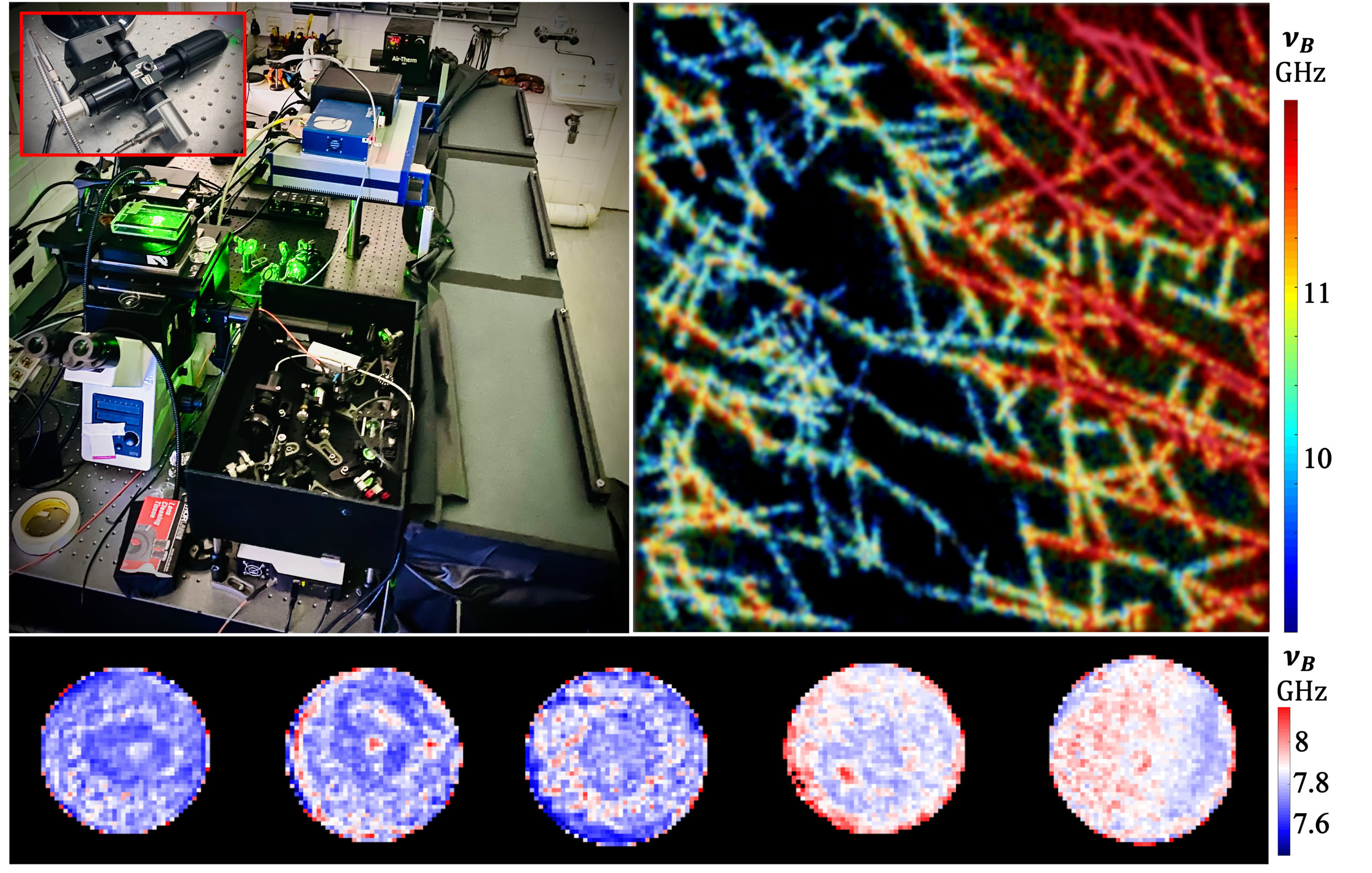
3. Angle-resolved Spontaneous Brillouin Light Scattering.
Custom inverted geometry. Parallel detection from multiple scattering angles. Excitation and detection NA tunable via spatial masks . Scan range: up to 500 microns (piezo stage) and optionally 2.5 cm (motor stage). Operating spectroscopy wavelength: 532nm (also changeable to 660nm by replacing several optical components). Elastic suppression via gas absorption cell (for 532nm) and spatial masks. Stable temperature control. Free Spectral Range: 30 GHz (15GHz or 60GHz possible by replacing several optical components). Suitable for extracting information on GHz-frequency mechanical anisotropy of reasonably transparent samples.

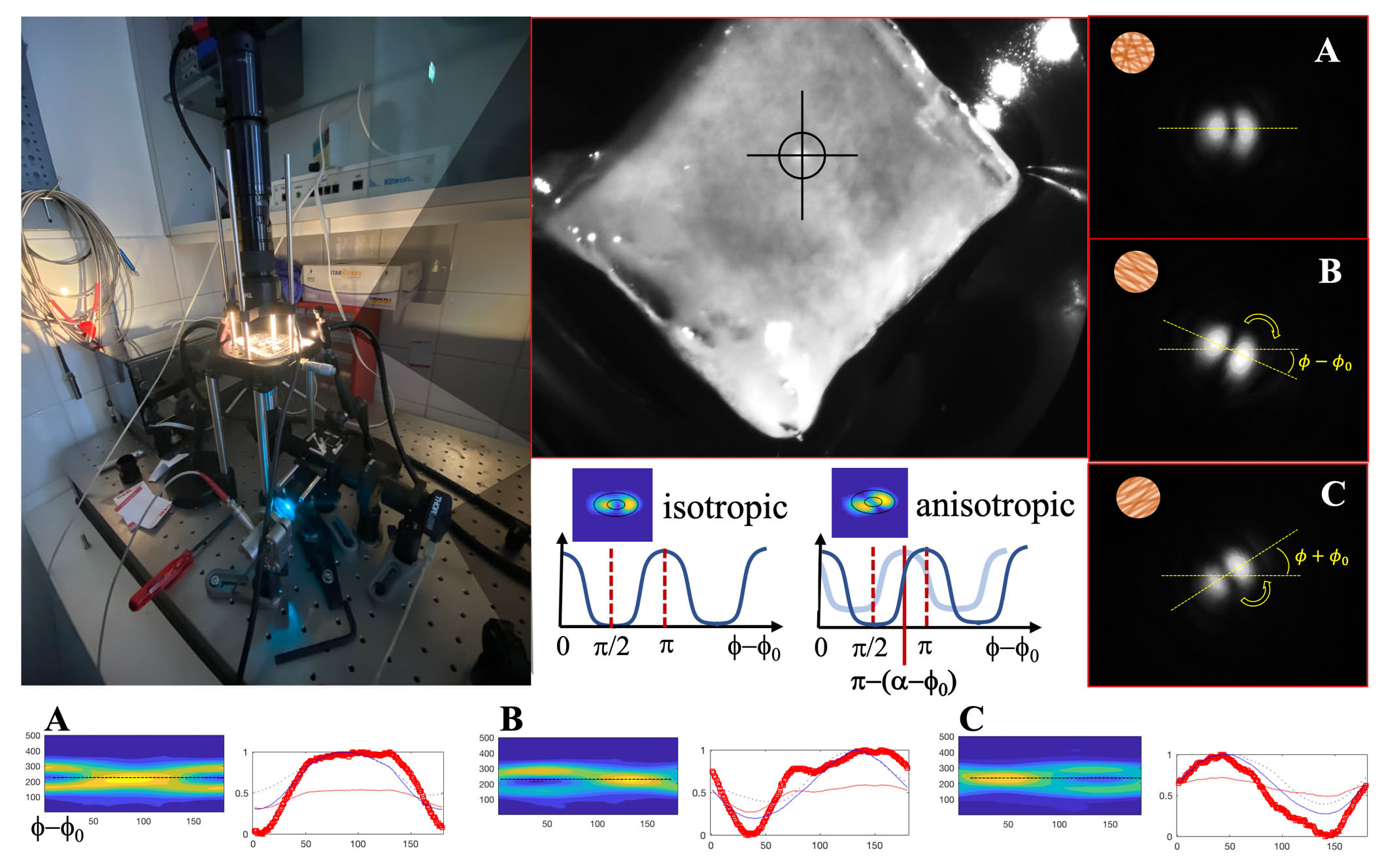
4. Polarization base measurements of birefringence/structural anisotropy.
Using polarized (491nm) light, structural anisotropy (orientational asymmetry) of samples can be extracted using a sensitive S-plate based polarization-resolved projection on a detector array. Effective numerical aperture <0.5, and adjustable via iris. Point measurements at discrete locations. Suitable for reasonably transparent samples.
5. Time-resolved imaging microspectroscopy.
For diverse applications that involve studying molecular and collective-molecular dynamic on time scales from ns-sec using line/area illumination and parallel 2D and 3D detection schemes. Back-scattering or off-axis (90o) detection. Excitation/detection: 0.1-1.49 NA. Scan range: up to 300 microns (piezo). Wavelengths: 405nm, 488nm, 514nm, 561nm, 642nm. Possible spectrally selective detection in 2D via band pass filters and 1D via grating or VIPA/etalon.