
High Resolution Episcopic Microscopy
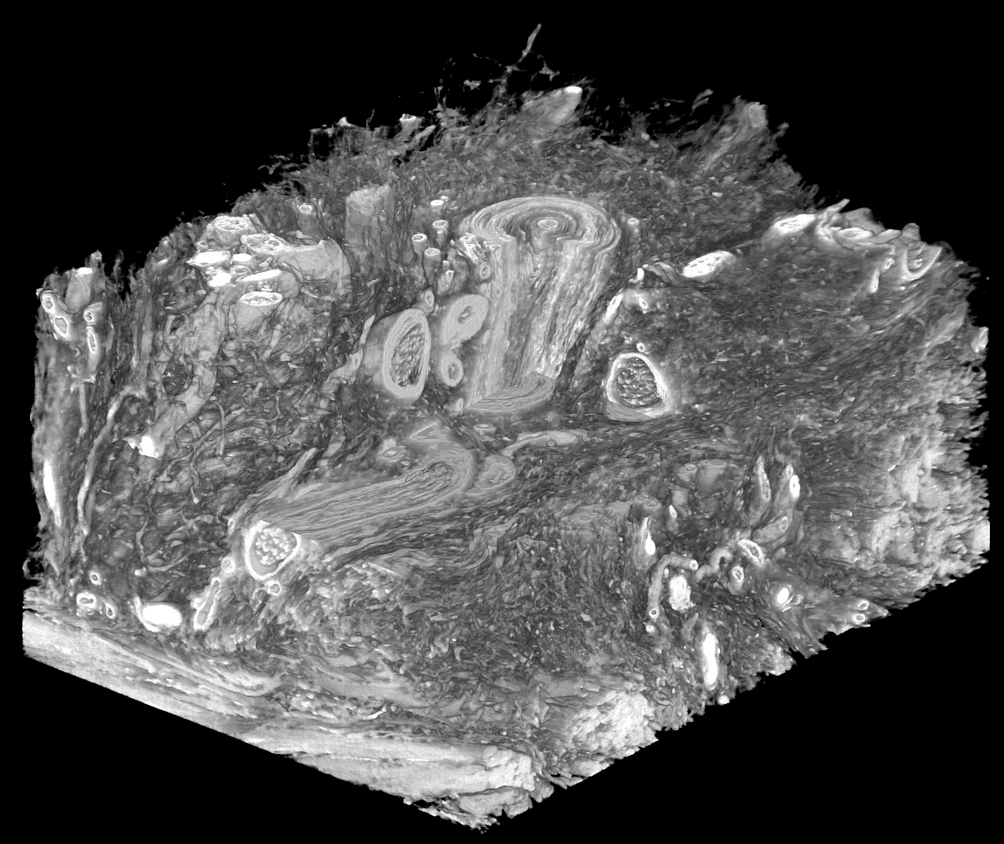
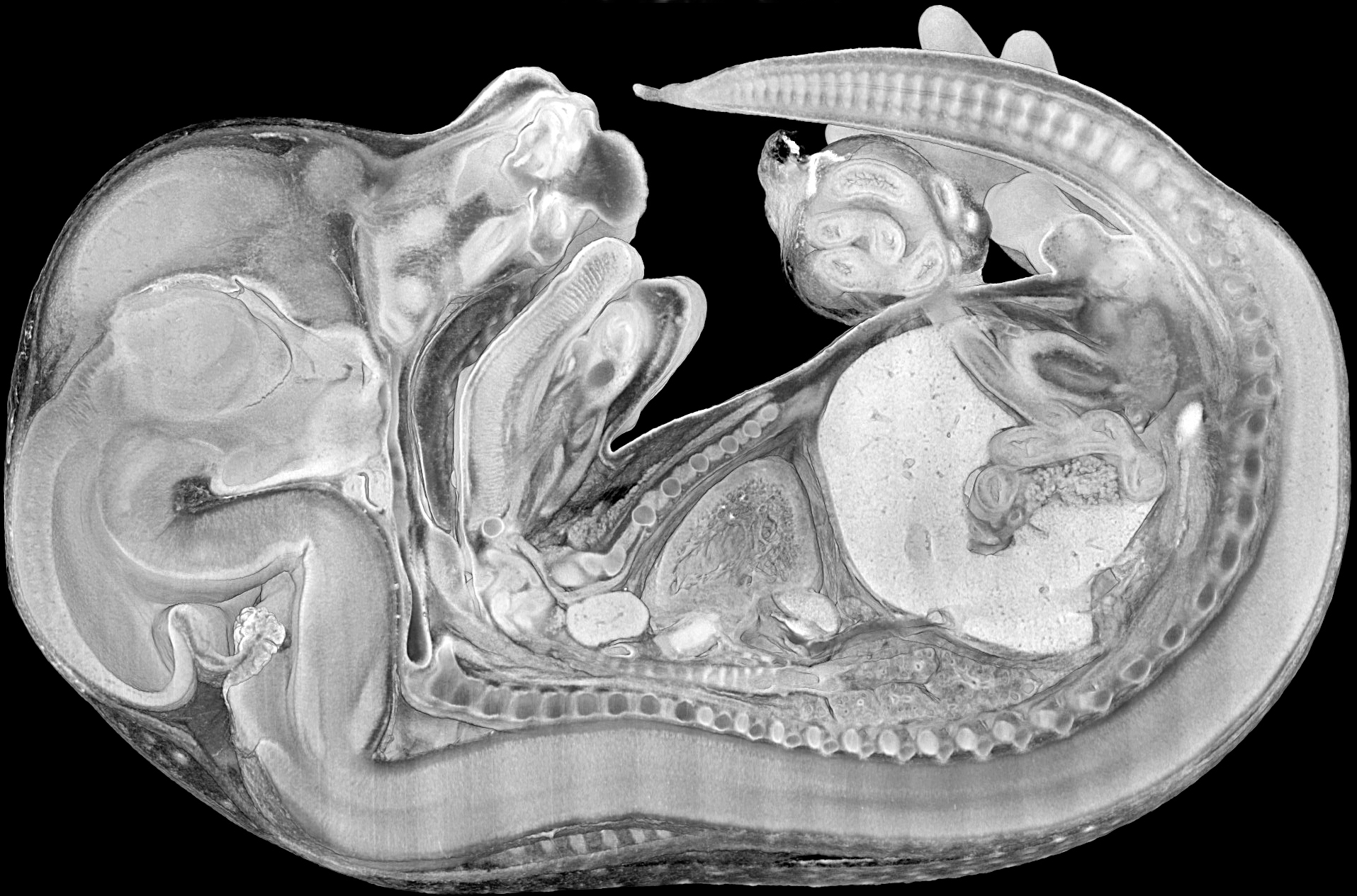
High resolution episcopic microscopy (HREM) is an ex-vivo imaging method, that generates three-dimensional volume data sets consisting of stacks of inherently aligned images. HREM works with fixated and histologically processed specimens, which are embedded in methacrylate resin and physically sectioned, while images are captured directly from the block surface. HREM data sets offer highly detailed grey-scale images of near histologic quality showing the overall morphology of a wide range of biological specimens. Volumes of up to 10 x 10 x 15 mm3 can be visualised with typical voxel sizes ranging from 1 x 1 x 1 µm3 to 6 x 6 x 6 µm3.
Resources
-
HREM apparatuses
-
High-end workstations
-
Software (Amira©, Osirix©) for visualisation and data analysis
Applications
-
Biomedical model organisms (mouse, chick, …)
-
Human tissue samples
-
Organic material