
Electron Microscopy Lab
The EM-lab of the Center for Anatomy and Cell Biology offers a wide range of preparation and imaging methods for examining ultrastructures of tissues and cells.
The imaging of biological samples is realized by transmission electron microscopy (TEM) and TEM tomography. Depending on the size and type of the sample, conventional chemical fixation, microwave fixation, as well as cryofixation can be used.
Both internal and external services are available on a collaborative basis. For an individual project design and information on usage fees, please contact us (Dr. Andy Sombke).
Equipment
Transmissionelectronmicroscopic imaging provides two-dimensional images. After embedding in resin, ultra-thin sections (40-50 nm) are prepared which electrons can penetrate. Alternatively, 3-dimensional imaging can be achieved by stepwise rotation of a thicker sample (TEM tomography).
The Transmission Electron Microscope Tecnai G20 can visualize objects up to 100,000 x magnification (higher magnifications dependent on specimen quality) with an accelerating voltage of 20-200 kV.
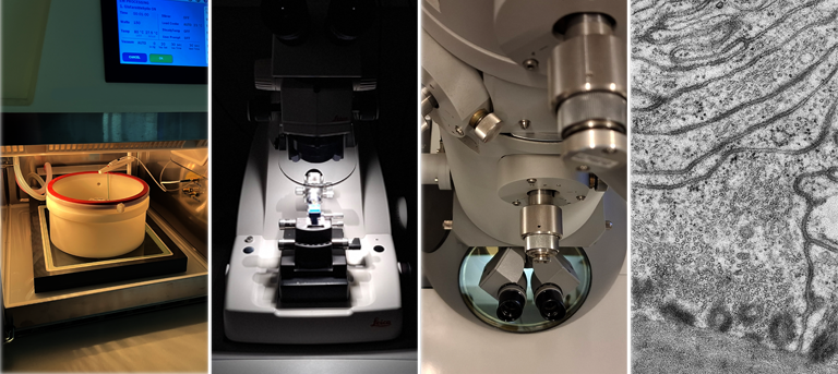
Fixation and sample processing:
- Leica EM AFS1 (Baltec HPM-010) for cryofixation
- Pelco BioWave Pro+ fixation microwave
- Leica ACE 200 for carbon coating
- Leica Ultratrim high-speed milling system
- Leica Ultracut UCT and 7 for semi- and ultra-thin microtomy
- Leica Ultracut 7 with Leica EM FC7 for cryo-microtomy
Imaging:
- 200 kV Tecnai G2 20 (FEI) transmission electron microscope with TEM-tomography option (with a FEI Eagle 4K CCD camera) for imaging and acquisition
- Zeiss AxioImager for comparative light-microscopic analyses
Established protocols:
- conventional chemical fixation of tissue samples, organoids, 3D cell culture samples and cell monolayers
- high pressure freezing and freeze substitution for high quality cell preservation
- negative staining for visualization of e.g. macromolecules and bacteria
- semi- and ultra-thin microtomy of resin-embedded specimens
- 3D reconstruction with Amira (ThermoFisher)