
AG Funktionelle Ultrastruktur
In the dynamic landscape of cell biology, understanding the complex details of functional ultrastructures is crucial for unraveling cellular processes. Our primary objective is to provide a comprehensive understanding of cellular dynamics by employing electron microscopic techniques to explore tissues and cells.
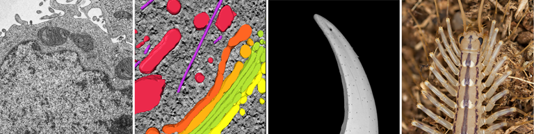
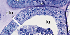
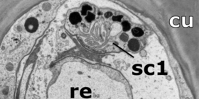
In addition to collaborative biomedical projects, our research explores various facets of arthropod anatomy, including the cellular organization and variability of epidermal and venom glands, the anatomy of respiratory systems, appendage development and regeneration, and the intricacies of sensory organs and primary processing centers within the nervous system. Centipedes exhibit venomous forcipules and evolutionarily innovative appendages, with the last pair of legs being particularly transformed in many species. These ultimate legs serve diverse functions like claw-like structures used for physical defense, sensory appendages resembling reverse antenna or aggregates of epidermal glands that transform the appendage into a predominantly glandular organ.
These insights not only contribute to our fundamental biological knowledge but also hold promising applications in the biomedical field. To analyze centipede anatomy, morphology, evolutionary transformations, and functional ultrastructures, we employ multimodal microscopic techniques, with a special emphasis on transmission electron microscopy.
Maurstad MF, Ramiro IBL, Oeyen JP, Sombke A, Büsse S, Nachtigall PG, Jakobsen KS, Undheim EAB (2026) The green lacewing venom system and the complex mechanisms underlying its evolution. Molecular Biology and Evolution 43(1): msaf326.
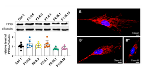
Valentin K, Kustermann M, Schneider MR, Aminfar H, Vollnhofer K, Wedrich A, Stapf C, Bertich M, Ritter M, Mendrina T, Valcanover D, Berger W, Eckhard M, Sombke A, Lilja SV, Paquay A, Rosensteiner B, Schmidt I, Bittner RE, Georgi TP, Pemp B, Schmidt WM (2026) A recurrent missense variant in the PPIB gene encoding peptidylprolyl isomerase B underlies adult-onset autosomal dominant optic atrophy. Genetics in Medicine 28(1): 101595..
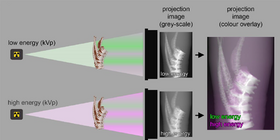
Handschuh S, Reichart U, Kummer S, Gabner S, Ruthensteiner B, Sombke A, Schwaha T, Beisser CJ, Lemell P, Haberthür D, Hlushchuk R, Glueckert R, Metscher BD, Glösmann M (2025) Microscopic dual-energy computed tomography (microDECT) imaging of animal tissues: the colour of X-rays. Methods in Microscopy 2: 249-301.
Zrelski MM, Eckhard M, Fichtinger P, Hösele S, Kustermann M, Sombke A, Mill L, Schmidt WM, Norwood F, Schlötzer-Schrehardt U, Wiche G, Schröder R, Winter L (2025) Impaired autophagic flux in skeletal muscle of plectin-related epidermolysis bullosa simplex with muscular dystrophy. Journal of Cachexia, Sarcopenia and Muscle 16 (4): e70001.
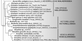
Waeschenbach A, Zhoua Z, Schwaha T, Okamura B, Gordon DP, Wood TS, Ostrovsky AN, Todd JA, Decker SH, Johnson M, Al-Ghanem MM, De Blauwe H, Florence WK, Graham R, Hall A, Hartikainen H, Jenkins HL, Kuklińskim P, Porter JS, Smith AM, Spencer Jones ME (2025) A genome skimming phylogeny of ctenostome bryozoans. Zoological Journal of the Linnean Society 204: zlaf060.
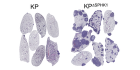
Luca AC, Kurnaeva M, John DK, Machtinger M, Vollmer NHJ, Moedl B, Hannich JT, Eckhard M, Lam HS, Musiejovsky L, Trenk C, Homolya M, Fürnsinn C, Sombke A, Schabbauer G, Eferl R, Sharif O, Casanova E, Mol HP (2025) Loss of SPHK1 fuels inflammation to drive KRAS-mutated lung adenocarcinoma. Cancer Letters 623: 217733.
Radhouani M, Farhat A, Hakobyan A, Zahalka S, Pimenov L, Fokina A, Hladik A, Lakovits K, Brösamlen J, Dvorak V, Nunes N, Zech A, Idzko M, Krausgruber T, Köhl J, Uluckan O, Kovarik J, Hoehlig K, Vater A, Eckhard M, Sombke A, Fortelny N, Menche J, Knapp S, Starkl P (2025) Eosinophil innate immune memory after bacterial skin infection promotes allergic lung inflammation. Science Immunology 10: eadp6231.
Barutia I & Sombke A (2024) Explosive regeneration and anamorphic development of legs in the house centipede Scutigera coleoptrata. Frontiers in Zoology 21:23.
Mosca E, Federa A, Pirker C, Schosserer M, Liendl L, Eckhard M, Sombke A, Dömötör O, Kirchhofer D, Timelthaler G, Baier D, Gurschka P, Gabler L, Reithofer M, Chin JM, Elsayad K, Englinger B, Tahir A, Kowol CR, Berger WS (2024) The tyrosine kinase inhibitor Nintedanib induces lysosomal dysfunctionality: role of protonation-dependent crystallization processes. Chemico-Biological Interactions 111243.
Züger S, Krings W, Gorb SN, Büscher TH, Sombke A (2024) Material composition and mechanical properties of the venom-injecting forcipules in centipedes. Frontiers in Zoology 21: 21.
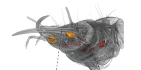
Schendel V, Müller CHG, Kenning M, Maxwell M, Jenner RA, Undheim EAB, Sombke A (2024) The venom and telopodal defence systems of the centipede Lithobius forficatus are functionally convergent serial homologues. BMC Biology 22: 135.
Mitić BM, Jovanović, VB, Todosijević MM, Eckhard M, Vasiljević LC, Tešević VV, Vujisić LV (2024) Chemical defence of a centipede (Clinopodes flavidus). Journal of Insect Physiology 155: 104649.
Decker SH, Saadi A, Baranyi C, Hirose M, Lemer S, Sombke A, Aguilera-Muñoz F, Vieira L, Smith A, Waeschenbach A, Schwaha T (2024) Boring systematics: a genome skimmed phylogeny of ctenostome bryozoans and their endolithic family Penetrantiidae with the desciption of one new species. Ecology and Evolution 14(4): e11276.
Neumüller J, Jungbauer C, Wagner T (2024) Characterization and Enumeration of Platelet Microvesicles in Human Platelet Concentrates by Using Transmission Electron Microscopy Including Electron Tomography. In: Huerta-Cuellar G (Ed.) Electron Microscopes and Their Applications.
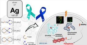
Teixeria RG, Stefanelli A, Pilon A, Warmers R, Fontrodona X, Romero I, Costa PJ, Villa de Brito MJ, Hudec X, Pirker C, Türck S, Antunes A, Kowol CR, Ott I, Brozovic A, Sombke A, Eckhard M, Tomaz AI, Heffeter P, Valente A (2024) Paraptotic cell death as unprecedented mode of action observed for new bipyridine-silver(I) compounds bearing phosphane co-ligands. Journal of Medicinal Chemistry 67(8): 6081-6098.